Giemsa Stain Kit (May-Grunwald)
Details
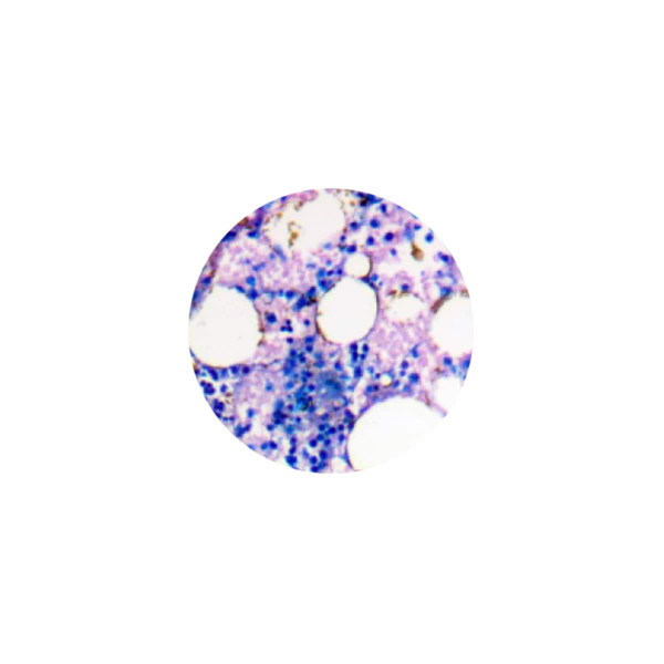
The Giemsa Stain Kit (May-Grunwald) is intended for use in the visualization of cells present in hematopoietic tissues and certain microorganisms. This kit may be used on formalin-fixed, paraffin-embedded or frozen sections.
Nuclei: Blue/Violet
Cytoplasm: Light Blue
Collagen: Pale Pink
Muscle Fibers: Pale Pink
Erythrocytes: Gray, Yellow, Pink
Rickettsia: Reddish-Purple
Helicobacter Pylori: Blue
Mast Cells: Dark Blue with Red Granules
Uses/Limitations:
- For In-Vitro Diagnostic use only
- Histological applications
- Do not use past expiration date
- Use caution when handling these reagents
Control Tissue:
- Blood Film
- Any well fixed tissue
Preparation of Reagents Prior to Beginning:
1. Prepare Working May-Grunwald Solution by mixing 25ml of May-Grunwald Solution with 25ml of Phosphate
Buffer Solution, pH 6.8.
2. Prepare Working Giemsa Solution by mixing 2.5ml of Giemsa Stock Solution with 50ml of Phosphate Buffer
Solution, pH 6.8.
Procedure (Standard):
1. Deparaffinize sections if necessary and hydrate to distilled water.
2. Place slide in staining tray and flood with Working May-Grunwald Solution for 6 minutes. Note: Agitate slide occasionally to insure proper staining.
3. Flood slide with Phosphate Buffer Solution, pH 6.8 until no stain runs off.
4. Flood slide with Working Giemsa Solution for 13 minutes. Note: Agitate slide occasionally to insure proper staining.
5. Flood slide with Phosphate Buffer Solution, pH 6.8 until no stain runs off.
6. Allow slide to remain in Phosphate Buffer Solution, pH 6.8 for an additional 3 minutes.
7. Dip slide quickly in distilled water and air dry at room temperature.
8. Dip slide in Xylene or Xylene Substitute.
9. Mount in synthetic resin.
Procedure (Mast Cells):
1. Deparaffinize sections if necessary and hydrate to distilled water.
2. Place slide in staining tray and flood with Working May-Grunwald Solution for 6 minutes. Note: Agitate slide occasionally to insure proper staining.
3. Flood slide with Phosphate Buffer Solution, pH 6.8 until no stain runs off.
4. Flood slide with Working Giemsa Solution for 13 minutes. Note: Agitate slide occasionally to insure proper staining.
5. Flood slide with Phosphate Buffer Solution, pH 6.8 until no stain runs off.
6. Differentiate by dipping slide in Acetic Acid Solution (0.25%) until background is desired intensity.
7. Dip slide 20 times in Phosphate Buffer Solution, pH 6.8.
8. Dip slide quickly in distilled water and air dry at room temperature.
9. Dip slide in Xylene or Xylene Substitute.
10. Mount in synthetic resin.
Click for Instructions for Use
| Components | Size | SDS Links |
|---|---|---|
| May-Grunwald Solution | 500 mL | View SDS |
| Giemsa Stock Solution | 500 mL | View SDS |
| Phosphate Buffer Solution, pH 6.8 | 500 mL |
Ratings & Reviews
No reviews available
Be the first to Write a Review